Parameters
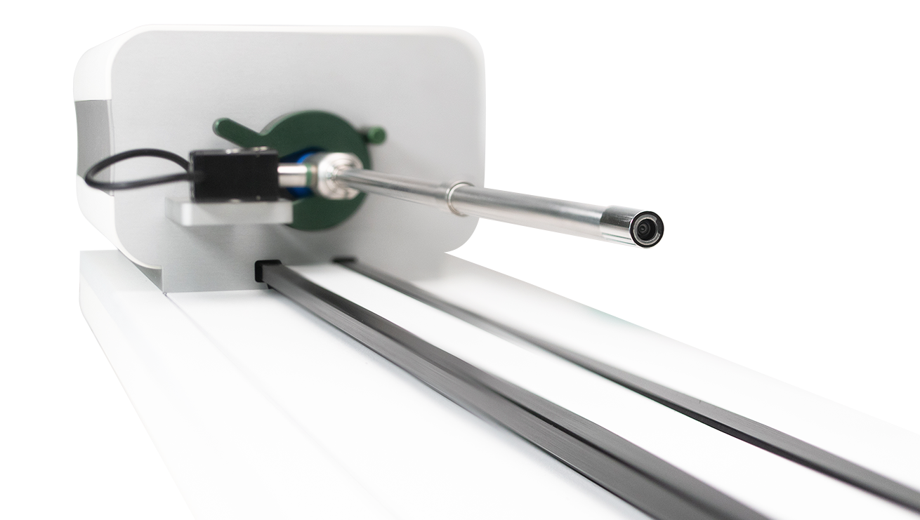
Rigid Endoscope Testing
LightControl and ScopeControl test rigid endoscopes on specific parameters. On this page you will find a clear explanation on how every measurement is done per parameter.
Need more info? NHS Case Study.
Clinical data? Here.
Light Transmission
Measures light transmission of the lenses (lux). The device uses a calibrated light source which passes through the endoscope to the eye piece which is connected to a camera.
The light intensity is calculated by detecting the visible area and isolating the white pixels. Then the amount of light is calculated from the image. Multiple shots with various shutter times are used to get histograms that are converted to determine a regression line with Y values. Every scope is measured with the same calculated Y value. The shutter times tells something about the LT. These values are compared with the photodiode and temperature sensor in the markersphere and corrected if needed.
The camera takes single consecutive images and the system records these at pixel level. Each Pixel has an RGB value in the color spectrum. The combination of all pixels is converted into a luminance value by an algorithm specifically developed for this task.
The system uses these images to cross-reference the measured value with data known about this brand and type of endoscope in the system.
Based on the measured value and the reference value, a score between 0 and 100% is given.
Focus
To check that the lenses are intact and not dirty.
This measurement is done to ensure a clear image. It prevents out-of-focus endoscopes from entering the operating room. The measurement is based on scientific research on blur detection for digital images using Harr Wavelet transformation. This method can quickly and accurately determine whether an image is blurry and to what extent an image is blurry. It is effective against both blur and linear motion blur.
In addition, its effectiveness is not affected by backgrounds in images.
The camera looks through the endoscope at a grid that shows "sharp" markers. During the measurement, the system takes several images to form a picture of the status of each lense.
The system uses an average reference value of a group of new endoscopes and gives a score from 0 to 100% for this specific endoscope.
The captured images are stored in endoscopemanager and are displayed in the report.
Light Fibers
Measures the light transmission of the fiber package in a rigid endoscope.
This measurement is done to ensure that the fibers in the endoscope allow enough light to pass through. This prevents an endoscope from having to be replaced during surgery because the image is not clear enough. If this parameter is too low, the light cone is too weak.
Light is emitted from the markersphere through the fibers to the light inlet of the light endoscope. A photodiode is attached, which captures the emitted calibrated LED light. Based on an original reference and reference from a group of new endoscopes, it is determined how much light passes through. This assessment essentially indicates how many fibers or what part of the fiber package no longer allows light to pass through.
This assessment is assigned a value from 0 to 100% for this specific endoscope based on an average reference value from a group of new scopes and it refers to previous measurements. It takes the best measured value as a new reference.
Color Correctness
Measures the discoloration of the rod lenses also known as the color balance. This measurement is done to see discoloration due to the sterilization process. It ensures quality in your process and alerts you when discoloration will cause misinterpretation of the image. It ensures patient safety.
Next to the Light transmittance data, the device takes images and measures shifts in color transmission (sRGB space) using the HSV model. HSV stands for Hue, Saturation, Value and describes color. Since the description is done in three dimensions, we speak of a color space.
The three components are determined independently and together determine the color: Hue is what we usually call "color", like a dot on the rainbow. In the HSV model, the color is plotted on a circle, and the spot is indicated in degrees: Hue thus runs from 0 to 360. Saturation indicates the brightness of a color. It is expressed in percentages and goes from 0% (pale, gray) to 100% (full color). Value of Brightness represents the lightness of the color. It is expressed in percentages and goes from 0% (black) to 100% (white).
The device converts the images of the RGB camera to the HSV model and cross-references them with the data from its selftest upon startup and with an average value of a group of new endoscopes known for this brand and type of endoscope in the system.
Based on the measured value and the reference value, a score between 0 and 100% is given.
Lens Fracture
Measures whether one or more internal lenses have a fracture.
This measurement is done to ensure that defective endoscopes with internal lens breakage do not enter the operating room, that endoscopes are repaired or replaced in a timely manner, and that endoscopes do not cause dangerous situations in patients.
The camera takes multiple images and makes a histogram of them. The system then references the image set to previous images of this brand anf type of endoscope and feeds this data into the neural network of endoscope manager. Based on this, a pass/fail is triggered. Following this, the images are added to the neural network.
Particle Detection
Measures whether particles have entered the internal sections of the endoscope.
This measurement is done to ensure that contaminated endoscopes do not enter the operating room, that endoscopes are repaired or replaced in a timely manner, and that endoscopes do not cause cross contamination.
The camera takes multiple images and creates a histogram. The system then refers the image set to previous images of this endoscope and feeds the data into the neural network of endoscope manager. Based on this, a pass/fail is triggered. The images are then added to the neural network.
Field of View
Measures the apparent maximum view of the objective lens.
This measurement is made to ensure that the image transmitted by the endoscope is wide enough to generate an adequate view. It measures the maximum field of view from the tip of the lens.
The camera is pointed at the sphere and looks through the endoscope at a calibrated matrix on a sphere and determines from this image how large the field of view is based on the dots displayed on the image. The system then counts these visible markers and converts this to the number of degrees in visibility.
After that the measurement is referenced to previous measurements and calculates the value based on a group of new endoscopes and any previous measurements of this particular endoscope.
In the case of a new, unknown endoscope, this measurement is entered as the initial reference in the endoscope manager system.
Viewing Angle
Measures the viewing angle of the endoscope.
This measurement is made to verify that the endoscope is still in spec. When it is out of spec, it indicates a dirty lense or lenses which are not placed apart correctly due to repairs.
The camera is pointed at the sphere and looks through the endoscope at red symbols. From this image it determines the viewing angle which is aimed at the center point inside the Sphere Assembly.
After that, the measurement is referenced to previous results and a value is calculated based on a group of new endoscopes and any previous measurements of this particular brand and type of endoscope.
In the case of a new, unknown endoscope, this measurement is entered as the initial reference in endoscope manager. Since, endoscopemanager consists of many endoscopes this does not happen very often.

